Contents: 2024 | 2023 | 2022 | 2021 | 2020 | 2019 | 2018 | 2017 | 2016 | 2015 | 2014 | 2013 | 2012 | 2011 | 2010 | 2009 | 2008 | 2007 | 2006 | 2005 | 2004 | 2003 | 2002 | 2001
2004, 12
Automatic retiming method based on genetic algorithm for the detection and the follow-up of dental lesions
language: English
received 16.08.2004, published 27.09.2004
Download article (PDF, 270 kb, ZIP), use browser command "Save Target As..."
To read this document you need Adobe Acrobat © Reader software, which is simple to use and available at no cost. Use version 4.0 or higher. You can download software from Adobe site (http://www.adobe.com/).
ABSTRACT
In dental surgery, a great number of pathologies (cysts, granuloma…) presents clinical, radiological and evolutionary aspects considerably polymorphic. Medical imagery and particularly oral imagery, by the means of the digitized images and the image analysis algorithms, constitutes an essential element which leads to a precise presumptive diagnosis. The technique of retiming per subtraction can be thus used to recognize in two-dimensional images evolution of the pathological zones (lesions, tumours). From this point of view, a method of automatic retiming was developed by genetic algorithm to follow the evolution of the pathological zones after parodental treatment. In this article the traditional technique of retiming by specifying its disadvantages for our application and then an automatic model of retiming, which offers promising results, are presented. This tool will make it possible to distinguish, identify, and visualize automatically the form of the pathological structures and their evolution in time. An analysis of form and texture of these structures will allow the identification type of pathology.
13 pages, 9 figures
Сitation: M. R. Benbrahim, R. Benslimane, El. Aalloula. Automatic retiming method based on genetic algorithm for the detection and the follow-up of dental lesions. Electronic Journal “Technical Acoustics”, http://www.ejta.org, 2004, 12.
REFERENCES
[1] E. Bonnet, J. L. Coudret, I. Fillere et al. Imagerie buccodentaire. Encyclopedie Medico Chirurgicale (Paris) 22-010-D-15. 1990.
[2] L. Daudibetieres, G. Etienne et al. Possibilites de traitement et d’analyses numeriques des cliches radiographiques dento-maxillaires. In Ondo-stomatologiques review. N°175. 1991.
[3] Bragger U, Burgin W, Fourmousis I, Schmid G, Schild U, Lang N. P. Computer assisted densitometric image analysis of digital substraction images: In vivo error of the method and effect of thresholding. Journal of Periodontology. 1999.
[4] Wenzel A, Warrer K, Karring T. Digital subtraction radiography in assessing bone changes in periodontal defects following guided tissue regeneration. J. Clin. Periodontol. 1992. Mar. 19(3). 208–13.
[5] Danesh-Meyer M. J., Chen S. T., Rams T. E. Digital subtraction radiographic analysis of GTR in human intrabony defects. Int. J. Periodontics Restorative Dent. 2002. Oct. 22(5). p. 441.
[6] M. K. Jeffcoat, M. S. Reddy, H. R. Van Der Berg, E. Bertens. Quantitative digital subtraction raddiography for assessement of peri-implant bone change. Clin. Oral Implant Res. 1992. N°3. 22–27.
[7] Allen K. M., Hausman E. Analytical methodology in quantitative digital subtraction radiography: analyses of the aluminum reference wedge. J. Periodontol. 1996. Dec. 67(12). 1317–21.
[8] Nummikoski P. V., Steffensen B., Hamilton K. et al. Clinical validation of a new subtraction radiography technique for periodontal bone loss detection. J. Periodontol. 2000. Apr. 71(4). 598–605.
[9] M. S. Reddy, J. M. Bruch, M. K. Jeffcoat, R. C. Williams. Contrast enhancement as an aid to interpretation in digital subtraction radiography. Oral Surg. Oral. Med. Oral Patho. 1991. N°71. 763–769.
[10] Bragger U, Burgin W, Marconi M et al. Influence of contrast enhancement and pseudocolor transformation on the diagnosis with digital subtraction images (DSI).
J. Periodontal Res. 1994. Mar. 29(2). 95–102.
[11] H. Grondahl, T. Okano, R. L. Webber. Staistical contrast enhancement of subtraction images for radiographic caries diagnosis. Oral Surg. Oral. Med. Oral Patho. 1982. N°53. 219–223.
[12] P. F.Van der Stelt, S. M. Dunn, A. Mol. Observer performance in digital subtraction radiography using non linear contrast enhancement. I. Dent. Res. 1993. 353–72.
[13] Bragger U., Fourmousis I., Burgin W. Standard Operating Procedures (SOP) for Computer Assisted Densitometric Image Analysis (CADIA) of Digital Subtraction Images (DSI) to reveal periodontal and periimplant tissue changes.
http://www.molar.unibe.ch/research/CADIA.
[14] D. E. Goldberg. Genetic Algorithm in Search, Optimization, and Machine Learning. Addison Wesley, 1989.
[15] Z. Michalewiez. Genetic Algorithms + Data Structure = Evolution Programs. Springer, Berlin, 1992.
[16] M. Buck and N. Diehl. Motion analysis and image sequence processing. Chapter “Model-based image sequence coding”. Kluwer Academic Publishers. 1993. 285–315.
[17] E. Dubois and J. Konrad. Estimation of 2-D motion fields from image sequences with application to motion-compensated processing. In Motion Analysis and Image Sequence Processing (M. Sezan and R. Lagendijk, eds.). Kluwer Academic Publishers, 1993. ch. 3, 53–87.
[18] J. M. Gorce, D. Friboulet, I. E. Magnin. Methode d'estimation du mouvement des parois cardiaques a partir d'images 3D. Innov. Techn. Biol. Med. 1994. Vol. 15. N°5.
[19] Declerck J., Feldmar J., Goris M. L., Betting E. Automatic registration and alignment on a template of cardiac stress and rest reoriented SPECT images. IEEE Transactions on Medical Imaging. 1997. 16. 727–37.
[20] C. Jerian, R. Jain. Determining motion parameters for scenes with translation and rotation. IEEE Trans. Pattern Anal. Machine Intell. 1984. Vol. 6. N°4.
[21] A. Ouaazizi, R. Benslimane. Motion estimation of images sequences by genetic algorithms. Inter. Conf. On IEEE Systems Machines and Cybernetics, Japan, 1999.
[22] T. E. Davis, C. J. Principe. A simulated annealing like convergence theory for the simple genetic algorithm. In Belez and Booker, 1991, 174–181.
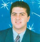 |
Benbrahim Med Reda was born in Fes, Morocco. He received the D.U.T diploma from High School of Technology of the Sidi Mohamed Ben Abdellah University, Fes, Morocco in 1990, the Master degree from University of Bordeaux, France in 1992 and the Ph.D. degrees from the University of Bordeaux, France in 1996. Since 1996, he is Assistant Professor at the High School of Technology, University of Sidi Mohamed Ben Abdellah (Fes, Morocco) and researcher member of the Laboratory of Transmission and Image Processing (LTTI). His research is focused on Medical Image Analysis. e-mail: r_benbrahim(at)yahoo.fr |
|
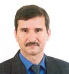 |
Rachid Benslimane received his Ph.D. from the University of Montpellier (France) in 1985. From 1986 to 1992 he has been an Assistant Professor in the University Sidi Mohamed Ben Abdellah, where he has received in 1992 his second Doctor's degree. From this date he was appointed Professor at the High School of Technology where he established the Laboratory of Transmission and Image Processing (LTTI). He supervised more than 15 Ph.D. thesis and managed research projects supported by National Institutions as well as European Projects. R. Benslimane has co-authored more than 80 papers including 17 journal papers. His scientific work deals with Image Processing and analysis, Image retrieval by content-based approach and data analysis. |
|
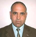 |
El Houssaine Aalloula - Head of Orthodontie Service of CHU IBN SINA, Vice-senior of the dental Faculty of Medicine, University Mohammed V. Suissi (Rabat, Morocco). His research is focused on dental pathologies and computer analysis. |
|